
Charting the Cranium pt 2
the CNS and the dicephalon

"The brain is like a muscle... Understanding is joyous." — Carl Sagan
Hi all! Back again talk about the brain!! This article may bounce around a little bit as I’m going to finish talking about the forebrain and also about the nervous system (as GSCE Biology was so long ago). Still, I hope to teach you something new about the brain!
As you probably know, the nervous system consists of two main parts: the central nervous system and the peripheral nervous system. The central nervous system (CNS) consists of the brain and spinal cord whilst the peripheral nervous system (PNS) contains the cranial nerves, spinal nerves, and peripheral ganglia (all the sweet extra stuff).
The CNS
Every part of the CNS is encased in bone, the spinal cord is supported within the vertebral column whilst the brain is the most protected organ in the body, covered by the skull. However this is not the brains only bodyguard. Additional protection (and rather uncomfortable fact) results from the brain floating in a pool of cerebrospinal fluid (CSF), this is to protect the neurons at the bottom of the brain from becoming squished (a term called neuronal buoyancy) and for shock absorption.
There is also a chemical barrier, known as the “blood brain barrier”, which is a highly selective, semipermeable cellular border that separates circulating blood from the brain’s extracellular fluid. This is because the brain requires a large supply of blood, continuously receiving approximately 20 percent of the blood flow from the heart. Whilst blood flow quantity varies to other parts of the body, depending on their needs and relative to those of other regions, the brain always receives its share. This is because the brain can only store small amounts of its fuel (typically glucose), meaning a consistent blood supply is essential. A simple one-second interruption of the blood flow to the brain uses up much of the dissolved oxygen, a six-second interruption produces unconsciousness and with blood flow cut off, after a few minutes blood flow, permanent brain damage results.
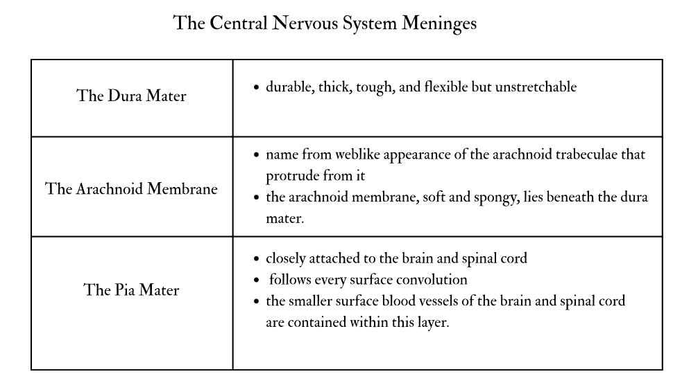
This is the introduction to a sexy little thing known as “Meninges”, a protective sheath that covers the central nervous system. The name comes from the Greek word for membrane, and it consists of three layers.
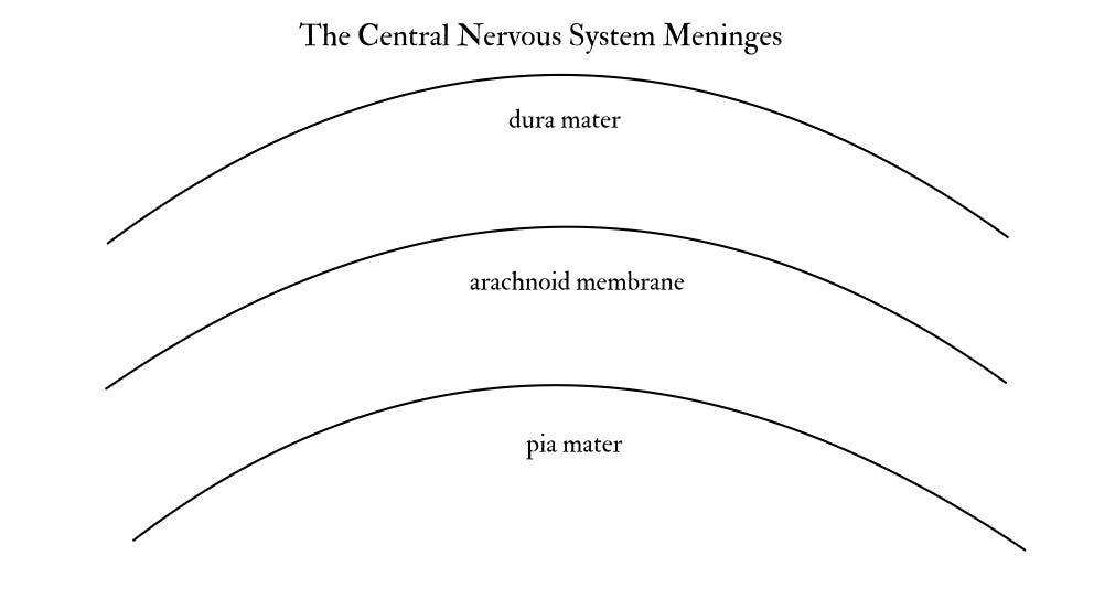
Between the pia mater and arachnoid membrane is a gap called the subarachnoid space, which is filled with cerebrospinal fluid (CSF).
Meanwhile the peripheral nervous system is covered with two layers of meninges. The middle layer (the arachnoid membrane), and its pool of CSF, covers only the brain and spinal cord. Instead, the outer and inner layers (dura mater and pia mater) fuse together and form a sheath that covers the spinal and cranial nerves and the peripheral ganglia.
Now we’ve got that cleared up: onto the ventricles !
The ventricular system of the brain consists of a series of hollow, interconnected chambers called ventricles, which are filled with CSF. The largest of these chambers are the lateral ventricles (or the first and second ventricles), which are connected to the third ventricle, which is located at the midline of the brain (The deep groove that separates the brain into the left and right hemispheres). The third ventricles walls divide the surrounding part of the brain into symmetrical halves. A bridge of neural tissue called the massa intermedia crosses through the middle of the third ventricle, whilst a long tube, the cerebral aqueduct, connects the third ventricle to the fourth ventricle.
The ventricles serve the very important function of producing and containing CSF. It is made by a special tissue with a rich blood supply called the choroid plexus, which extends into all four of the ventricles.

As the brain is very jellylike and soft. The weight of a human brain (approximately 1.4 kg), along with its fragile construction, means that it is of upmost importance that it be protected from shock. This problem is resolved by its immersion in CSF, whereby the brain’s net weight is reduced to only approximately 80 g, thereby the pressure on the base of the brain is considerably reduced. The CSF surrounding the brain and spinal cord also helps reduce the shock to the CNS that would be caused by sudden head movement.
Now, back to the forebrain!
As I mentioned last time, the brain is VERY complex. Last week we covered the telencephalon, so this week we’ll cover the rest of the forebrain, the diencephalon. This is situated between the telencephalon (the cerebral cortex and its subcortical structure) and the mesencephalon (the midbrain); and it surrounds the third ventricle (see I told you it was important).
Whilst the diencephalon consists of 4 structures (the Thalamus, Hypothalamus, Epithalamus, Subthalamus), we’ll only go in depth on the first 2 and the pineal gland (located in the Epithalamus).

The thalamus makes up the dorsal part of the diencephalon. It sits near the middle of the cerebral hemispheres, and is immediately medial and caudal to the basal ganglia. It has two lobes, which are connected by the massa intermedia (much like the third ventricle).
Most of the neural input to the cerebral cortex is firstly received from the thalamus; insofar as much of the cortical surface being divided into regions that receive projections from specific parts of the thalamus.
The thalamus consists of several nuclei, with some thalamic nuclei receiving sensory information from the sensory systems. The neurons in these nuclei then relay the sensory information to specific sensory projection areas of the cerebral cortex (i.e. the lateral geniculate nucleus receives information from the eye and sends axons to the primary visual cortex, whilst the medial geniculate nucleus receives information from the inner ear and sends axons to the primary auditory cortex). However, other thalamic nuclei that project to specific regions of the cerebral cortex, do not relay sensory information (i.e. the ventrolateral nucleus receives information from the cerebellum and projects it to the primary motor cortex). Still other nuclei receive information from one region of the cerebral cortex and relay it to another region, for example, several nuclei help to control the general excitability of the cerebral cortex, therefore, they projection in a widespread manner to all cortical regions.
Hypothalamus
The hypothalamus is located underneath the thalamus, and whilst it is relatively small structure, it’s very important. The hypothalamus’ role relates to controlling the autonomic nervous system and the endocrine system, and organising behaviours related to the survival of the species, such as fighting, escape, eating, drinking, sleeping and reproduction.
It is situated on both sides of the ventral portion of the third ventricle and is a complex structure, containing many nuclei. Additionally, much of the endocrine system is controlled by hormones produced by cells in the hypothalamus, a special system of blood vessels directly connects the hypothalamus with the anterior pituitary gland.
The pituitary gland is attached to the base of the hypothalamus via the pituitary stalk, meanwhile in front of the pituitary stalk is the optic chiasm, where half of the axons in the optic nerves (from the eyes) cross from one side of the brain to the other.
The hypothalamic hormones are secreted by specialised neurons called neurosecretory cells, located near the base of the pituitary stalk. These hormones job is to stimulate the anterior pituitary gland to secrete its hormones. Most of these hormones secreted control over other endocrine glands, i.e. the gonadotropic hormones stimulate the gonads to release male or female sex hormones, which affect cells throughout the body, including some in the brain. Though other anterior pituitary hormones, such as the somatotropic hormone (the growth hormone)—do not control other glands and rather act as a final messenger.
The hypothalamus also produces and controls the secretion of the hormones of the posterior pituitary gland, oxytocin and vasopressin. Oxytocin and vasopressin are involved in a number of different physiological and behavioural functions, including pair bonding and parental behaviour.
And finally: The pineal gland. This is even smaller than the hypothalamus and is tucked inbetween the two lobes of the thalamus. Its primary purpose is producing the hormone melatonin, which, as you most likely know, regulates our sleep-wake cycles. However, it is also important for regulating hormonal functions, like the hypothalamus.
Thanks for reading!